Geautomatiseerde levende cel beeldvorming
Cel Biologie - Kankercellen - Stamcellen - Ontdekking van geneesmiddelen
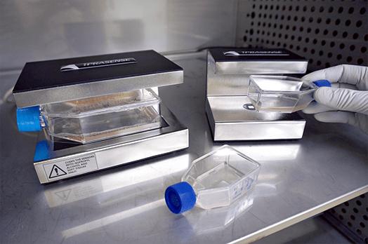
Nieuwe perspectieven voor live celbeeldvorming en celkinetische analyse. De labelvrije time-lapse beeldvormingstechnologie biedt een veelzijdige oplossing voor het monitoren van celkweek in uw incubator. Het ongeëvenaarde extra grote gezichtsveld en de afwezigheid van focus voor het uitvoeren van robuuste real-time analyse van aanhangende cellen in elke container (petrischaal, T-Flask, objectglaasje...).
Het CYTONOTE productassortiment vereenvoudigt de techniek van live celbeeldvorming en transformeert de complexe en dure microscoop in een kosteneffectieve oplossing.

ETIKETVRIJ &
HOOG CONTRAST

ALTIJD
IN BEELD

INSTELLINGEN
GRATIS

GROOT GEZICHTSVELD
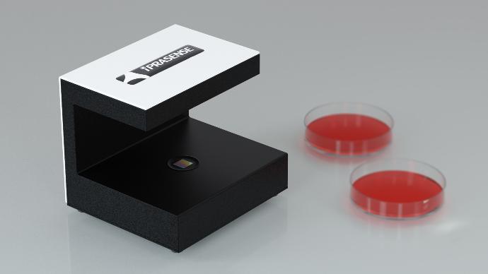
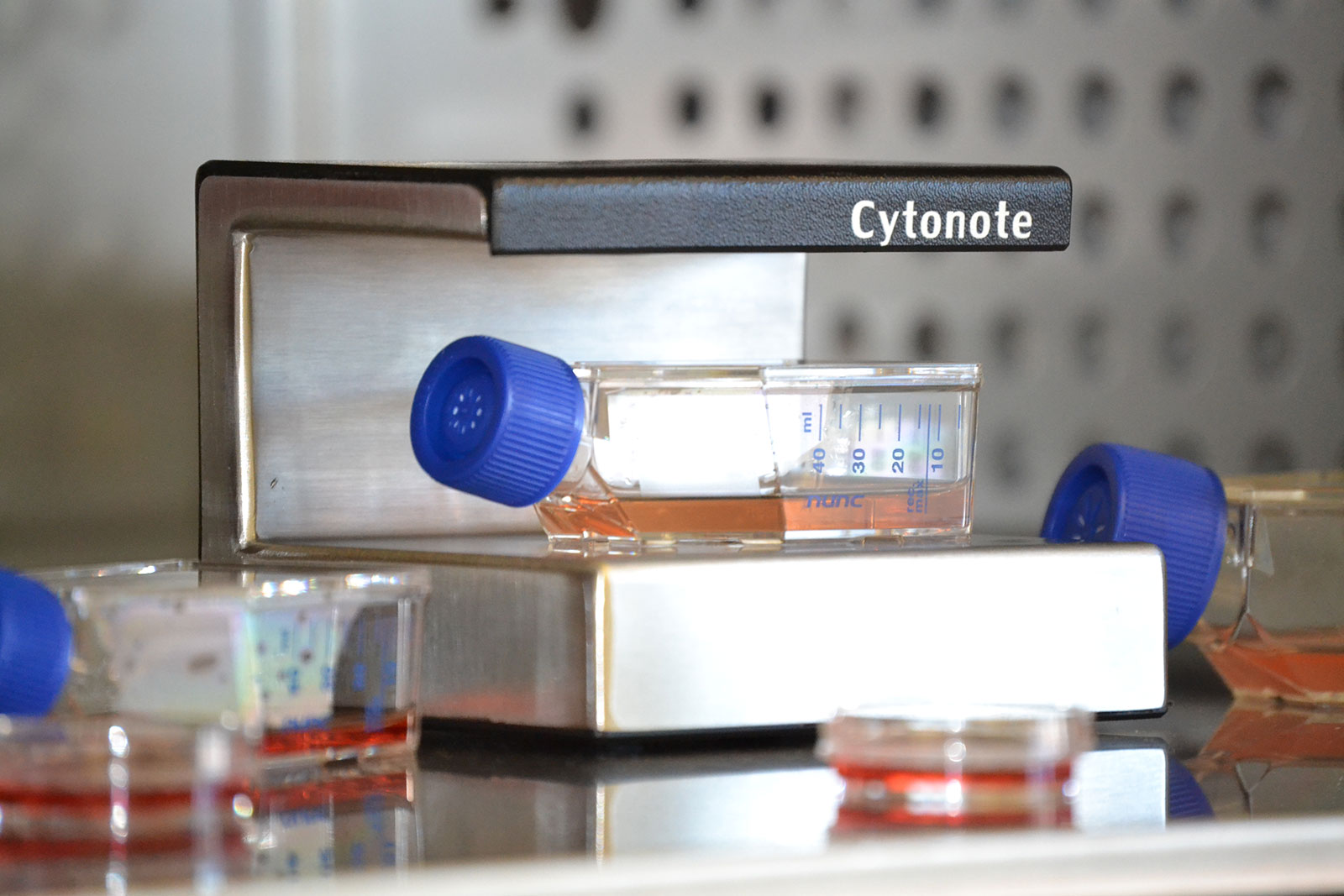
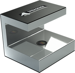
CYTONOTE 1 W
Met dit compacte apparaat kun je time-lapse en real-time beelden van je cellen verkrijgen vanuit je incubator.
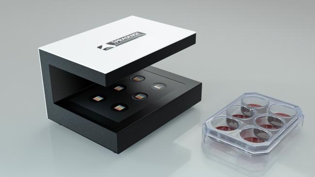
CYTONOTE 6 W
Ontworpen voor parallelle bewaking van celkweken in 6-wells platen. Hiermee kunt u time-lapse en real-time beelden van uw cellen verkrijgen vanuit uw incubator.
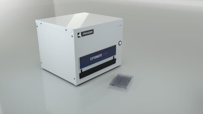
CYTONOTE SCAN
Ontworpen om time-lapse en real-time beelden van uw multiwellplaat te verkrijgen vanuit uw incubator.
Ontdek deze tijdsverloopsequentie van duizenden celdelingen!
De CYTONOTE is nu ongelooflijk met Phase Quantitative Imaging Capability en altijd betere beeldkwaliteit!
Een geweldig hulpmiddel voor celcyclusonderzoek product van IPRASENSE
Neem contact met ons op voor meer informatie of vraag een demo aan
Andere behoeften voor het bewaken van je celkweek?
Vraag het onze experts
Bekijk onze oplossingen voor geautomatiseerde telling en analyse van de levensvatbaarheid van cellen, analyse van metabolieten in kweekmedia, analyse door flowcytometrie, cellen sorteren, automatisering van pipetteerbewerkingenaseptische behandeling, wassen en oogsten van cellenenz.
Onze producten
Time-lapse imaging device - 1 Well Reader (1W), used for real time analysis of the cells in Petri dishes, T-flasks, slides or microfluidic chips from inside the incubator.
The CYTONOTE implements a patented Lensless Technology in a tiny device design to fit within any incubator.
The small foot print (132 cm² only!) allows you to follow any of your cell culture without any incubator adaptation or working space limitation.
Working on a ultra wide field of view, all data recovered from pictures taken by CYTONOTE 1W are representative of your cell culture. Indeed, working on 29.4mm² allows you to follow several thousand cells at a time.
Real-time Imaging cell culture allows you to browse through the whole acquisition set, play the timelapse, or export it into video format.
The HORUS Software associated with it is designed to monitor up to 6 Cytonotes simultaneously for 6 parallel or independent cell cultures.
Main Applications :
CELL MIGRATION
Chemotaxis, wound healing on high statistical number of cells and a very wide area.
CELL PROLIFERATION
Cell proliferation is determined through cell count and quantitative confluence determination. Growth Curve based on cell count or confluence for both adherent and non-adherent cells
ANGIOGENESIS
The very wide area allows for observing the full angiogenesis process with a high level of detail.

To use your Cytonote into a real-time imaging device inside your incubator

To transform your Cytonote into a real-time imaging device inside your incubator and allow connection of up to 6 Cytonote Reader on a single HORUS Software

Control Software for your Cytonote or Norma devices from Iprasense
