Types of imaging
- DNA
Gel Imaging:
DNA gel imaging involves capturing visual representations of separated DNA fragments on a gel through techniques like gel electrophoresis. Stained or labeled DNA is illuminated, using ethidium-bromide or safe dyes such as SYBR Green - Chemiluminescence
Imaging:
Chemiluminescence imaging is a method to visualize molecules, such as proteins or nucleic acids, by detecting the light emitted during a chemical reaction. This technique is often used in applications like Western blotting, providing sensitive and quantitative data on specific molecules. - Fluorescence
Imaging:
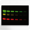
Fluorescence imaging utilizes fluorescent dyes or proteins to label target molecules, enabling their visualization under specific wavelengths of light. Widely employed in molecular and cellular biology, this technique allows for real-time tracking and localization of specific components within biological samples. - In-Vivo
Optical Imaging:
In-vivo optical imaging involves non-invasive visualization of biological structures or processes within a living organism using light. This technique, commonly used in medical research, provides real-time insights into physiological functions, disease progression, and the effects of treatments in a living system.
What solutions has Analis to offer?
Analis supplies imaging solutions from Vilber Lourmat.
VILBER is a leading life science company that develops and manufactures imaging and analyzing systems for fluorescence, chemiluminescence and bioluminescence applications. Vilber offers imaging solutions for gel documentation, western blot and in-vivo imaging of small animals and plants.
Contact us to discuss which instrument is the best fit
for your needs and budget


