Confocal Microscopy WORKSHOP
ANALIS ACADEMY WORKSHOP


CONFOCAL MICROSCOPY WORKSHOP
Join us to experience
the NEW Evident FV5000 microscope systems
Wednesday May 27, 2026
Thursday May 28, 2026
Monday June 1, 2026
Morning Workshop Sessions from 10 am to 12 am
Afternoon Workshop Sessions from 01 pm to 04 pm
FREE REGISTRATION
Schedule your session and if you prefer another time, simply indicate which one in the registration form
LOCATION
Eindhoven University of Technology
LCTE Microscopy Facility (room 0.410), Vector Building,
Dominee Theodor Fliednerstraat 2,
5631 BN Eindhoven, The Netherlands
Special thanks to Zyb Baster for hosting our Evident microscopy workshop session.
Microscopy Facility Manager of Cell and Tissue Engineering Laboratory at the Department of Biomedical Engineering at Eindhoven University of Technology
WORKSHOP SESSION
FV5000 confocal laser scanning microscope
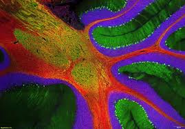
Experience the deepest imaging with FV5000.
Equipped with revolutionary
SilVIR detectors with the highest photon detection efficiency on the market in
visible and near infrared range. Coupled with ten lasers (including 785nm), the
sharpest full-field resonant scanner and patented spherical aberration
corrected volume imaging it is ideal for the sharp, deep and fast organoid
imaging. In addition we are bringing two single wavelength MPE lasers (920 and
1064 nm) with the possibility to go even deeper with second harmonic, green and
red labels.

ANALIS & EVIDENT SEMINAR - CONFOCAL LASER SCANNING MICROSCOPY
How FV5000/MEP make powerful imaging more accessible to capture more detail, more reliably in less time?
Eindhoven University of Technology
room 2.429, Vector Building
Dominee Theodor Fliednerstraat 2,
Wednesday May 27, 2026 - From 10.00 to 11.00 am
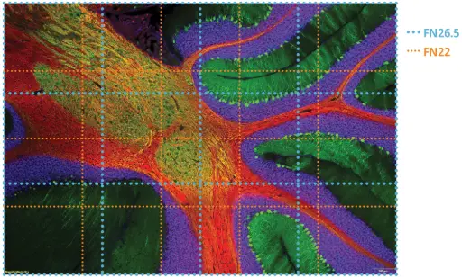
A Wider Window into Cellular Dynamics
Whether you're working with large, thick samples or multi-well plates, the IXplore IX85 SpinXL speeds up the process and enables you to quickly acquire massive quantities of data.
With an unparalleled 26.5 mm field number (FN), you can capture large tissues using 30% fewer images than FN22, and 15% fewer images than FN25.*
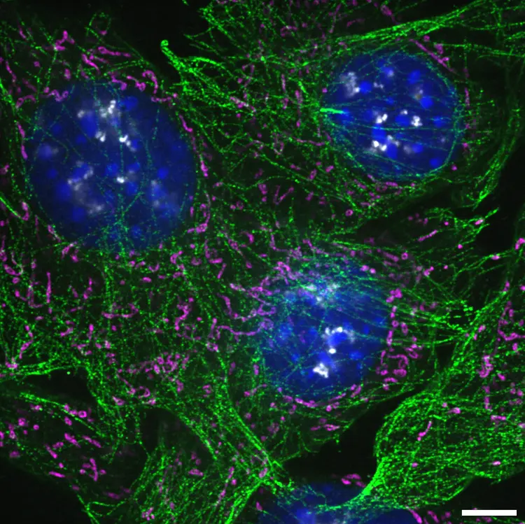
Super-Resolution Imaging
Spinning disk super-resolution systems equipped with optical photon reassignment (SoRa technology) deliver enhanced spatial detail while maintaining high acquisition speeds, achieving lateral resolution down to approximately 120 nm at up to 200 frames per second—about two times higher than standard spinning disk confocal imaging. This approach enables live-cell–compatible super-resolution imaging without requiring specialized sample preparation, helping researchers preserve physiological conditions. Typical applications include visualizing cytoskeletal dynamics, vesicle trafficking, and synaptic structures, where improved resolution and temporal performance are essential for capturing rapid subcellular events.
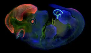
Developmental Biology
Spinning disk systems are well suited for embryo imaging, enabling long-term observation of developmental processes over hours or days with minimal disruption to normal growth. Gentle illumination helps reduce phototoxic effects, supporting healthy embryo development throughout extended time-lapse experiments. Multiposition acquisition enables researchers to track multiple specimens at the same time, improving experimental efficiency and statistical robustness. Common model organisms include zebrafish, Drosophila, C. elegans, and mouse embryos, where dynamic processes such as cell differentiation, morphogenesis, and tissue organization can be studied in real time.