High-Performance Imaging Systems for Advanced Life Science Research
Olympus / Evident system microscopes deliver cutting-edge imaging solutions for researchers needing high precision, speed, and flexibility. From macro imaging to confocal and super resolution, these platforms support complex live-cell and multi-dimensional studies.
Built for Advanced Applications:
- Exceptional image clarity – high resolution across macro to nano scale
- Broad system compatibility – fluorescence, confocal, super resolution
- Smart automation – faster workflows, reduced user handling
- Seamless software integration – with AI tools and 3D rendering options
Explore Our High-End Life Science Systems
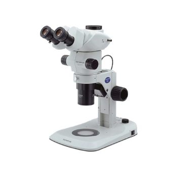
- Macro Zoom Microscopes – Zoom from whole organisms to subcellular detail with high contrast
- Super Resolution Microscopes – Break the diffraction limit for sharper, deeper cell imaging
- Confocal Laser Scanning Microscopes – Acquire clear 3D images from deep within tissues
Discover the right Evident system for your research. Analis provides local support, demos, and full system integration across Belgium, Luxembourg, and the Netherlands.
See all high-end products
Take your imaging and your discoveries further than ever before
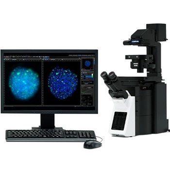
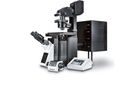
Evident is setting a new standard in life science imaging for neuroscience, cell biology, drug discovery, cancer research, and developmental biology with the launch of its new FLUOVIEW™ FV5000 confocal and multiphoton laser scanning microscope.

IXplore™ IX85 SpinXL and SpinSR spinning disk confocal microscopes expand research possibilities for imaging rapid cellular dynamics
whether your priority is high-throughput screening or super-resolution detail.
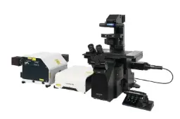
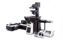
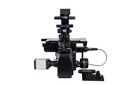
IXplore IX85 SpinXL Offers a Wider View into Cellular Dynamics
Powered by CrestOptics X-Light spinning disk technology, the IXplore IX85 SpinXL offers an unparalleled 26.5 mm field number (FN) that enables users to quickly capture cellular dynamics and rapidly screen large areas.
Learn more about IXplore IX85 SpinXL microscope
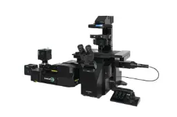
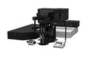
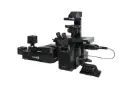
IXplore IX85 SpinSR Empowers Faster Discovery in
Stunning Detail
The IX85 SpinSR simplifies the capture of super-resolution details, empowering users to discover new insights faster.
Learn more about IXplore IX85 SpinSR microscope
Both the IX85 SpinXL and IX85 SpinSR microscopes can be paired with the world’s first silicone gel pad objective (LUPLAPO25XS) for accurate 3D volumetric reconstruction and deeper focus into samples.
Frequently asked questions
Here are some common questions about IXplore™ IX85 SpinXL and SpinSR spinning disk confocal microscopes.
Choose the right spinning disk system based on your imaging priorities: throughput or resolution.
SpinXL vs SpinSR - Quick Comparison
| SpinXL | SpinSR | |
| Primary strength | Large field imaging & throughput | Super-resolution & fine detail |
| Best for | High-content screening, large samples, multi-well plates | Subcellular structures, organoids, nanoscale dynamics |
| Field of view | Extra-large (26.5 mm) | Standard (optimized for resolution) |
| Resolution | Confocal resolution | Super-resolution (~120 nm) |
| Typical use | Fast population-level imaging | Detailed intracellular analysis |
Quick guidance:
- SpinXL = see more, faster
- SpinSR = see smaller, sharper
- Choose SpinXL if you need to image large areas quickly and increase throughput.
Ideal for screening workflows, multi-position experiments, and capturing dynamic processes across many cells simultaneously. - Choose SpinSR if you need to resolve fine cellular structures.
Ideal for studying subcellular organization, organoids, and nanoscale biological processes with enhanced detail.
Not sure which configuration is right for your workflow?
Our imaging specialists support you in selecting, configuring, and validating the right system for your applications.
Request a demo or application consultation
Our products


Key Advantages at a Glance
- Photon-level quantitative imaging with SilVIR™ cooled SiPM detectors
- Widest dynamic range (400–900 nm) for clear low-signal and high-signal imaging in one acquisition
- Dual scanning system:
- Resonant imaging up to 438 FPS (2048x32)
- Galvo scanning up to 8192×8192 px (8K) with 0.2 µs dwell time
- Super-resolution down to 120 nm (FV-OSR software, no extra hardware)
- Up to 6 simultaneous spectral channels with TruSpectral/VPH detection
- Fully integrated smart automation: AI sample search, laser optimization, shading correction, auto correction collar
- Multiphoton-ready platform with cost-effective fiber-pigtailed IR lasers or advanced tunable MPE systems
- Compatible with inverted, upright, gantry, and multiphoton frames
- Stable, reproducible performance with built-in Laser Power Monitor & Microscope Performance Monitor
Contact Analis application specialists to receive personalized system proposal or organize a demo.

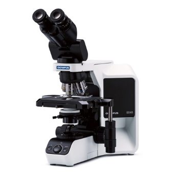
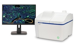
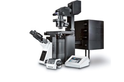
All-in-one benchtop fluorescence microscope designed for ease of use and high-quality imaging.
The Olympus / Evident APX100 streamlines fluorescence microscopy with smart automation and fast data access.
- Capture publication-grade images in just a few clicks
- AI-powered smart acquisition and processing
- Compact design for benchtop efficiency
- Integrated touchscreen and intuitive interface
- Ideal for labs with space or time constraints
Simplify your microscopy without sacrificing quality. Contact Analis for an on-site demo or configuration help.
- Compatible with most inverted fluorescence microscopes
- Ideal for life science research, neurobiology, membrane studies, cytoskeleton & large tissues
- Integrated NEO software for real-time acquisition, reconstruction, and quantification
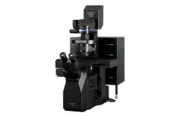
Key Advantages of SAFe Platform
- SAFe 180: Large 200 × 200 µm² FOV for high-density 2D nanoscopy using TIRF, HiLo, or EPI
- RedSTORM: Optimized 2D system for fast, high-precision red-channel STORM experiments
- SAFe 360: Combines DONALD & Magnified Astigmatism for full 3D SMLM with axial precision ~20–30 nm
- Real-time image reconstruction with drift correction and advanced quantification tools
- Multicolor capabilities: Sequential and spectral demixing modes
- AI-enhanced data interpretation via integrated analysis tools (Voronoi, DBSCAN, Ripley’s K, cluster metrics)
- TruSpectral technology allows simultaneous multiplexing of up to 6 channels
- Industry-leading 10-laser system (405–785 nm) with powerful NIR capabilities up to 900 nm
- Dual scanner system offers fast resonant and high-resolution galvo scanning with improved depth and speed
- SilVIR detectors provide stability and reproducibility across sessions
- Suitable for both fixed and live-cell imaging
Contact Analis application specialists to receive personalized system proposal or organize a demo.
- Powered by SilVIR™ detectors for low noise and high photon sensitivity
- TruResolution™ objectives minimize spherical aberration in deep tissue
- Resonant scanner enables high-speed, gentle time-lapse acquisition
- Modular system fits upright, inverted, or gantry configurations
- Wide spectral range with up to six channels and IR excitation from 690–1300nm
Ask Analis for a customized solution or live demo.
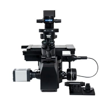
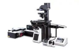
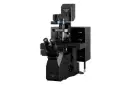
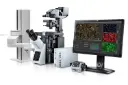
Inverted Microscope for Live-Cell Imaging and Automation
Motorized and Modular Design for Precision Imaging
Seamless integration with Olympus cellSens software ensures intuitive operation, automated workflows, and consistent image acquisition across experiments.
Flexible Configurations for Time-Lapse and High-Content Applications
- Supports fluorescence, DIC, phase contrast, and IR-DIC imaging
- Motorized Z-drive and XY stage with TruFocus™ Z-drift compensation
- Modular deck structure for easy integration of additional optical paths or accessories
- Seamless compatibility with incubation systems for live-cell viability
- Ideal for cancer biology, stem cell tracking, and neuroscience research
- Scalable for routine imaging to AI-assisted image acquisition and automated analysis
The IXplore IX85 platform is also available in two optimized configurations:
- IXplore IX85 Pro for high-speed, high-content imaging with a wide field of view
- IXplore IX85 Live for long-term live-cell imaging with minimized phototoxicity and precise environmental control.

Key Features & Benefits
- Super-resolution imaging down to 120 nm with dual microlens spinning disk
- High-speed acquisition up to 255 frames per second (full field)
- Live-cell compatibility with low phototoxicity and photobleaching
- Dual camera port & simultaneous multicolor acquisition
- TruFocus™ Z-drift compensation for stable long-term imaging
- Wide spectral range (400–750 nm excitation / 400–800 nm emission)
- Seamless integration with Olympus cellSens software
Key Features & Benefits
- Extra-large 26.5 mm FOV for fast, high-content acquisition
- Up to 498 fps imaging speed (full frame)
- NIR imaging capability reduces phototoxicity and improves depth
- Multi-disk flexibility: standard, thick sample, or high-throughput spiral
- TruFocus™ Z-drift compensation for multipoint time-lapse stability
- Compatible with AI-powered Olympus cellSens software
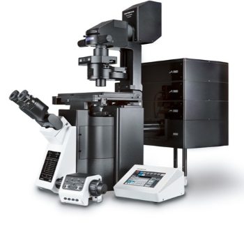
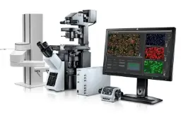
Key Features and Benefits
- Modular Olympus-based platform for assay development and high-throughput multiwell imaging
- Real-time cytometric analysis with multiparametric gating, classification, and object detection
- Integrated ScanR AI module for deep learning segmentation and rare event detection
- TruFocus™ IR laser autofocus for fast and precise z-positioning
- Supports advanced acquisition modes: Z-stack, time-lapse, multicolor, and label-free imaging
- Seamless integration with spinning disk confocal (CSU-W1), TIRF, and FRAP imaging modules
- Intuitive data visualization with real-time histograms, scatter plots, and kinetic tracking
- Enables end-to-end image acquisition, gating, cell isolation, and sequencing in one workflow.
- Compatible with laser microdissection and downstream RNA/DNA sequencing pipelines
- Adaptable to R&D labs, multiuser facilities, and clinical screening environments
Applications
- Cell viability, proliferation and apoptosis studies
- Live-cell kinetic and time-lapse tracking
- Gene expression, translocation, and reporter assays
- Cell cycle and nuclear morphology
- Drug screening and rare event detection
- Label-free fluorescence and low-exposure analysis
- FISH, PML body, comet assays, and multicolor fluorescence assays