Why Choose the KU-F40 Feces Analyzer for Parasitic Diagnostics?
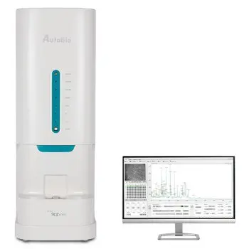
The KU-F40 Feces Analyzer, provided by our supplier Keyu Biological Engineering Co., Ltd., delivers a standardized and fully automated method for detecting and identifying parasites in fecal samples. This system combines the gold-standard microscopic approach with integrated AI for enhanced accuracy.

Easy to use - One step from the sample to the result.
New Special Sample Collection Cup: Features a rotating threaded screw cap for convenient sample collection.
Physical Detection: Automatically captures a photo of the sample and analyzes it using artificial intelligence to determine the physical characteristics of the feces.
Antigen Tests: A color image of the colloidal gold cartridge is captured by the camera for rapid detection. You can add up to six rapid tests to your fecal analysis: Rota/Adeno, H. Pylori, FOB, Calprotectin, and Lactoferrin.
Morphological Detection: Utilizes a microscope to enlarge fecal elements, combined with a high-definition camera for automatic image capture. The integration of digital imaging and artificial intelligence software technology ensures precise diagnostics.

Iodine Staining Function: Under Iodine staining mode, KU-F40 can automatically add iodine staining solution to stain the sample, improve the detection rate of special Ova and Parasite
Test Speed: 15 - 60 samples/hour
Microscope: High and low power objective lens with functions of auto focus upon startup and one-push auto focus
Camera: 5 mega-pixel HD CMOS camera
Image Quantity: More than 300 images, supporting multiple combinations and customized quantity of shooting fields
Image Capture Mode: Multi-field layered scanning up to 8 layers per field

Software Functionality

- One software platform to review all samples, designed for ease of use and clarity.
- The software enhances sample review with automatic parasite recognition and categorization of positive images to simplifying the validation process.
- Automatic identification of form elements :
| Blood | Egg of Ascaris lumbricoides | Blastocystis hominis |
| Calcium Oxalate Crystal | Egg of Clonorchis sinensis | Endolimax nana |
| Charcot Leyden Crystal | Egg of Diphyllobothrium latum | Entamoeba coli |
| Epithelial cell | Egg of Enterobius vermicularis | Entamoeba histolytica/dispar |
| Erythrocyte | Egg of Hookworm | Giardia lamblia |
| Fat Droplet | Egg of Hymenolepis nana | Other Amoeba |
| Fungus | Egg of Tapeworm | |
| Ganoderma Lucidum Spore | Egg of Trichuris Trichiura | |
| Leukocyte | Strongyloides stercoralis | |
| Macrophage | ||
| Muscle fiber | ||
| Plant cell | ||
| Pus Cell | ||
| Starch granule |