Skip to Content
EVIDENT

Abbelight SAFe Super-Resolution Nanoscopy Systems

Reference: EVI-Abbelight SAFe

https://www.analis.com/web/image/product.template/135195/image_1920?unique=689d05d
Modular Single-Molecule Nanoscopy from 2D to 3D with High Precision and Large FOV
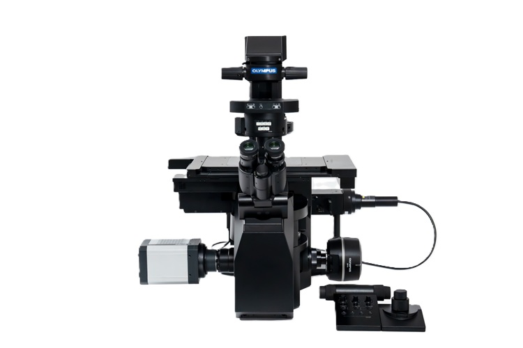
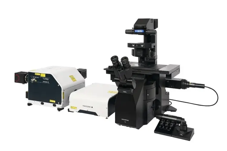
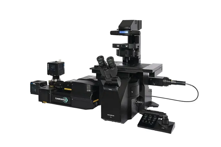
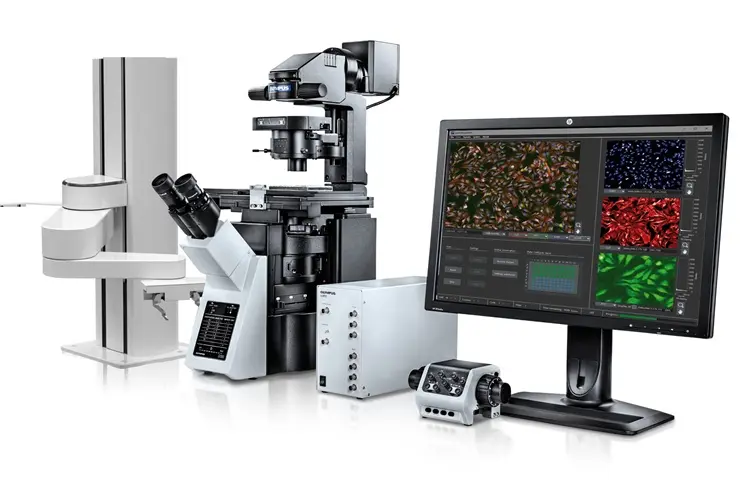
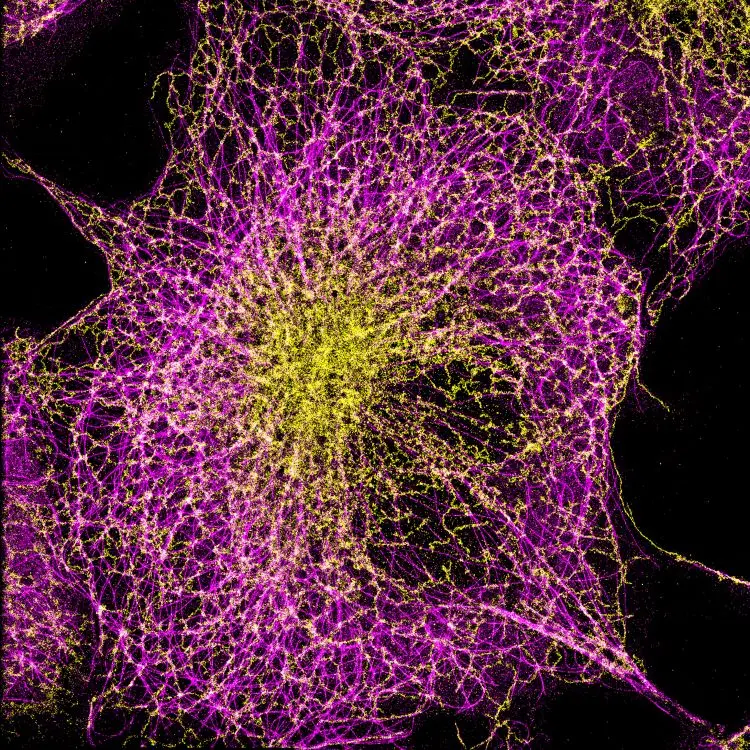
The Abbelight SAFe series brings powerful super-resolution microscopy to inverted fluorescence microscopes. These plug-and-play modules enable 2D and 3D Single-Molecule Localization Microscopy (SMLM) techniques like STORM, PALM, and PAINT, delivering nanometric resolution across a wide field of view. From basic 2D nanoscopy (SAFe 180), to optimized red-channel imaging (RedSTORM), up to full 3D axial and lateral localization (SAFe 360), the SAFe platform scales with your research.

  • Compatible with most inverted fluorescence microscopes
  • Ideal for life science research, neurobiology, membrane studies, cytoskeleton & large tissues
  • Integrated NEO software for real-time acquisition, reconstruction, and quantification

Key Advantages of SAFe Platform

  • SAFe 180: Large 200 × 200 µm² FOV for high-density 2D nanoscopy using TIRF, HiLo, or EPI
  • RedSTORM: Optimized 2D system for fast, high-precision red-channel STORM experiments
  • SAFe 360: Combines DONALD & Magnified Astigmatism for full 3D SMLM with axial precision ~20–30 nm
  • Real-time image reconstruction with drift correction and advanced quantification tools
  • Multicolor capabilities: Sequential and spectral demixing modes
  • AI-enhanced data interpretation via integrated analysis tools (Voronoi, DBSCAN, Ripley’s K, cluster metrics)

    This combination does not exist.

    Technical information

    Feature

    SAFe 180

    RedSTORM

    SAFe 360

    Imaging Mode

    2D SMLM (STORM, PALM, PAINT)

    2D Red-optimized STORM

    3D SMLM with DAISY (DONALD + Astigmatism)

    Field of View

    200 × 200 µm

    200 × 200 µm

    200 × 200 µm

    Z Resolution

    ~22–30 nm

    Z Stack Depth

    Up to 10 µm

    Multicolor Imaging

    Sequential

    Sequential (Red-optimized)

    Simultaneous (Spectral Demixing)

    Real-Time Reconstruction

    Yes

    Yes

    Yes

    Sample Types

    Fixed cells, dense labeling

    Membranes, cytoskeleton, actin-rich samples

    Thick tissues, neurons, multiplexed samples

    Drift Correction

    Yes

    Yes

    Yes (live correction via cross-correlation)

    Software

    NEO SAFe Suite (Acquisition + Analysis)

    NEO SAFe Suite

    NEO SAFe Suite (3D visualization + quant tools)

    Export Formats

    TIFF, CSV

    TIFF, CSV

    TIFF, CSV

    Download the brochure for full specifications of all modules.
    Contact Analis for expert guidance, configuration advice or a live demo.

    Related documents

    DOCUMENT NAME

    RELEASE DATE

    DOWNLOAD

    Brochure EVIDENT Abbelight SAFe Super-Resolution Nanoscopy Systems EN.pdf
    30 / 06 / 2025

    Related accessories, consumables & products

    IX85 Inverted Microscope System
    EVIDENT

    IX85 Inverted Microscope System

    Reference : EVI-IX85
    The IXplore IX85 is a high-performance, motorized inverted microscope platform designed for advanced live-cell imaging, time-lapse acquisition, and automated workflows in demanding life science applications.

    Inverted Microscope for Live-Cell Imaging and Automation

    Motorized and Modular Design for Precision Imaging

    Built on the proven Olympus IX3 frame and the legacy of the IX83 platform, the IXplore IX85 delivers enhanced automation, stability, and modularity to support a broad range of imaging techniques, from brightfield to fluorescence and DIC. It delivers precise control for long-term observations, high-content screening, and multi-position acquisition.
    Seamless integration with Olympus cellSens software ensures intuitive operation, automated workflows, and consistent image acquisition across experiments.

    Flexible Configurations for Time-Lapse and High-Content Applications

    • Supports fluorescence, DIC, phase contrast, and IR-DIC imaging
    • Motorized Z-drive and XY stage with TruFocus™ Z-drift compensation
    • Modular deck structure for easy integration of additional optical paths or accessories
    • Seamless compatibility with incubation systems for live-cell viability
    • Ideal for cancer biology, stem cell tracking, and neuroscience research
    • Scalable for routine imaging to AI-assisted image acquisition and automated analysis

    The IXplore IX85 platform is also available in two optimized configurations:

    • IXplore IX85 Pro for high-speed, high-content imaging with a wide field of view
    • IXplore IX85 Live for long-term live-cell imaging with minimized phototoxicity and precise environmental control.
     Contact Analis for expert guidance, a tailored configuration, or to book a live demo.
    scanR High-Content Screening Solution
    EVIDENT

    scanR High-Content Screening Solution

    Reference : EVI-scanR
    High-Content Imaging Cytometer for Automated Cellular Analysis in Life Science and Multiomics Workflows
    The ScanR system is a flexible, modular solution that combines the precision of an Olympus IX83 inverted microscope with the throughput and automation of a high-content screening platform. Designed for cell biology, drug discovery, systems biology, and live-cell assays, it delivers real-time image-based cytometry with AI-assisted segmentation, population gating, and deep learning analysis—ideal for demanding, high-throughput workflows.

    Key Features and Benefits

    • Modular Olympus-based platform for assay development and high-throughput multiwell imaging
    • Real-time cytometric analysis with multiparametric gating, classification, and object detection
    • Integrated ScanR AI module for deep learning segmentation and rare event detection
    • TruFocus™ IR laser autofocus for fast and precise z-positioning
    • Supports advanced acquisition modes: Z-stack, time-lapse, multicolor, and label-free imaging
    • Seamless integration with spinning disk confocal (CSU-W1), TIRF, and FRAP imaging modules
    • Intuitive data visualization with real-time histograms, scatter plots, and kinetic tracking
    • Enables end-to-end image acquisition, gating, cell isolation, and sequencing in one workflow.
    • Compatible with laser microdissection and downstream RNA/DNA sequencing pipelines
    • Adaptable to R&D labs, multiuser facilities, and clinical screening environments

    Applications

    • Cell viability, proliferation and apoptosis studies
    • Live-cell kinetic and time-lapse tracking
    • Gene expression, translocation, and reporter assays
    • Cell cycle and nuclear morphology
    • Drug screening and rare event detection
    • Label-free fluorescence and low-exposure analysis
    • FISH, PML body, comet assays, and multicolor fluorescence assays