EVIDENT
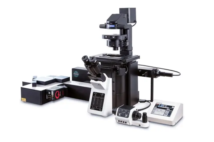
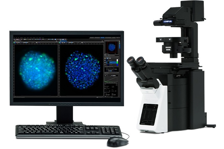
IX85 Inverted Microscope System
Reference: EVI-IX85
https://www.analis.com/shop/evi-ix85-ix85-inverted-microscope-system-129841 https://www.analis.com/web/image/product.template/129841/image_1920?unique=41bc2edThe IXplore IX85 is a high-performance, motorized inverted microscope platform designed for advanced live-cell imaging, time-lapse acquisition, and automated workflows in demanding life science applications.
Inverted Microscope for Live-Cell Imaging and Automation
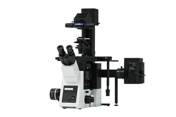
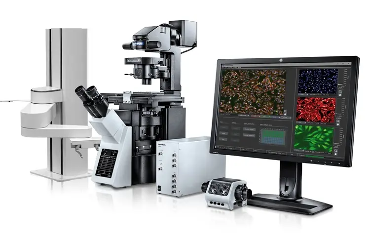
Motorized and Modular Design for Precision Imaging
Built on the proven Olympus IX3 frame and the legacy of the IX83 platform, the IXplore IX85 delivers enhanced automation, stability, and modularity to support a broad range of imaging techniques, from brightfield to fluorescence and DIC. It delivers precise control for long-term observations, high-content screening, and multi-position acquisition.
Seamless integration with Olympus cellSens software ensures intuitive operation, automated workflows, and consistent image acquisition across experiments.
Seamless integration with Olympus cellSens software ensures intuitive operation, automated workflows, and consistent image acquisition across experiments.
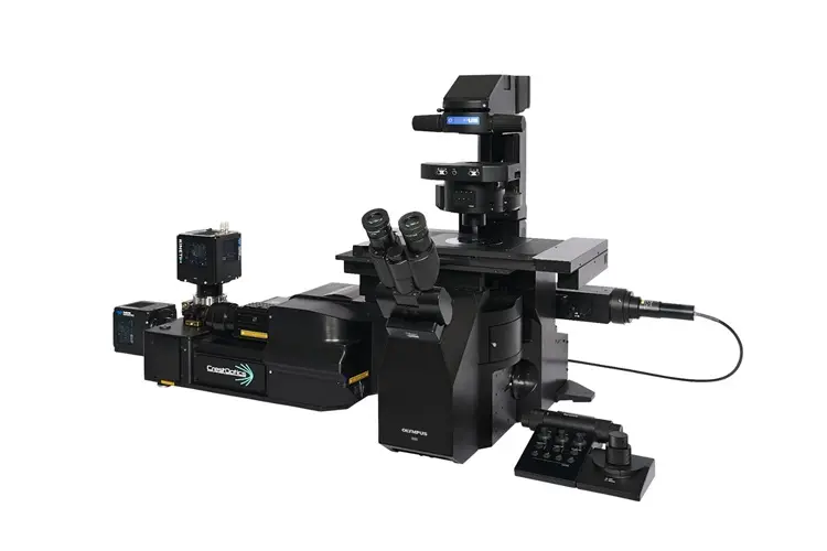
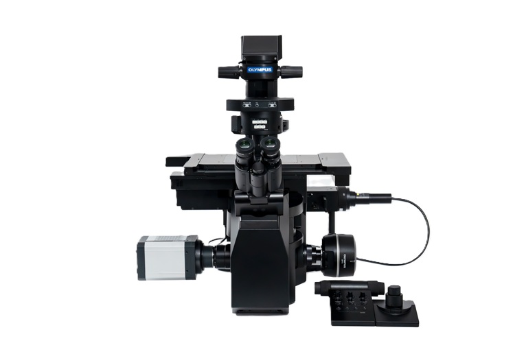
Flexible Configurations for Time-Lapse and High-Content Applications
- Supports fluorescence, DIC, phase contrast, and IR-DIC imaging
- Motorized Z-drive and XY stage with TruFocus™ Z-drift compensation
- Modular deck structure for easy integration of additional optical paths or accessories
- Seamless compatibility with incubation systems for live-cell viability
- Ideal for cancer biology, stem cell tracking, and neuroscience research
- Scalable for routine imaging to AI-assisted image acquisition and automated analysis
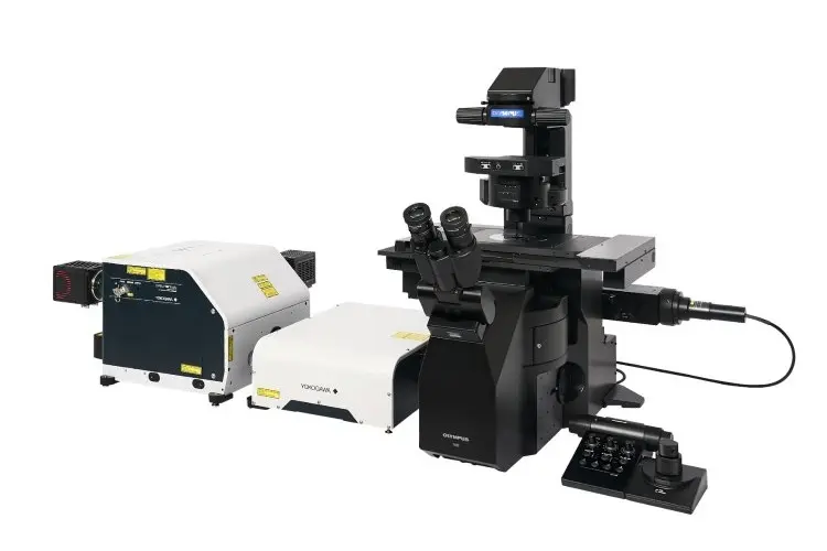
The IXplore IX85 platform is also available in two optimized configurations:
- IXplore IX85 Pro for high-speed, high-content imaging with a wide field of view
- IXplore IX85 Live for long-term live-cell imaging with minimized phototoxicity and precise environmental control.
Contact Analis for expert guidance, a tailored configuration, or to book a live demo.
Technical information
| Category | Specification |
| Microscope Frame | Olympus IX3-based, motorized IX85 with modular deck structure |
| Imaging Modes | Brightfield, Phase Contrast, DIC, Epifluorescence, IR-DIC |
| Automation | Motorized focus (Z), XY stage, shutters, filter wheels, and condenser |
| Stage Options | Encoded or encoded/motorized XY stage with multi-position control |
| Objectives | Supports 4X to 100X; universal turret (6 positions) |
| Camera Compatibility | Wide-field and sCMOS/high-speed camera port available |
| Live Cell Support | Incubator compatibility, vibration stability, IR-LED transmitted light |
| Dimensions (base frame) | Approx. 350 mm × 550 mm × 550 mm (H × W × D), frame only |
Download the full brochure for detailed system configurations.