Get Ready to Break the Boundaries of EV Detection — Go Deeper!
Extracellular vesicles (EVs) are key mediators of intercellular communication, influencing immune responses, tissue regeneration, tumor growth, and more. However, their analysis remains challenging due to the limitations of traditional cytometry in detecting EVs smaller than 100 nm.
With the CytoFLEX Nano, push the limits of detection and unlock new possibilities.
A New Approach to Nanoscale Flow Cytometry
The Beckman Coulter CytoFLEX Nano
Nanoscale flow cytometry is an advanced technique that integrates flow cytometry principles with nanotechnology. It enables the analysis of particles at the nanoscale, providing detailed information on size, composition, and surface properties.
This technology offers broad applications in biology, medicine, and materials science. It allows researchers to analyze and characterize nanoparticles, extracellular vesicles, and other small particles with exceptional precision and sensitivity.
Nanoscale flow cytometry also holds strong potential for developing diagnostic tools and targeted drug delivery systems. The ability to analyze nanoparticles based on their properties opens new avenues for personalized medicine and nanomedicine.
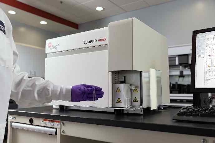
Detect Nanoparticles Down to 40 nm — With Multiparameter Fluorescence
The CytoFLEX Nano is the first purpose-built nanoscale flow cytometer capable of detecting nanoparticles—such as extracellular vesicles (EVs)—as small as 40 nm, while simultaneously performing multiparameter fluorescence detection.
Its software interface, CytExpert Nano, offers the sophistication required to explore the nanoscale range, while maintaining the ease of use that characterizes the CytoFLEX platform. As a result, answering complex research questions becomes easier than ever for EV researchers.
What Makes the CytoFLEX Nano Unique?
Built on the same principles as the CytoFLEX platform, the CytoFLEX Nano integrates the capabilities of multiple techniques into a single instrument. It enables:
- Detection of nanoparticles as small as 40 nm
- Analysis of individual EV markers, even at low expression levels (<10 antigen copies)
- Standardized, fast, and accurate analysis—without relying on multiple systems
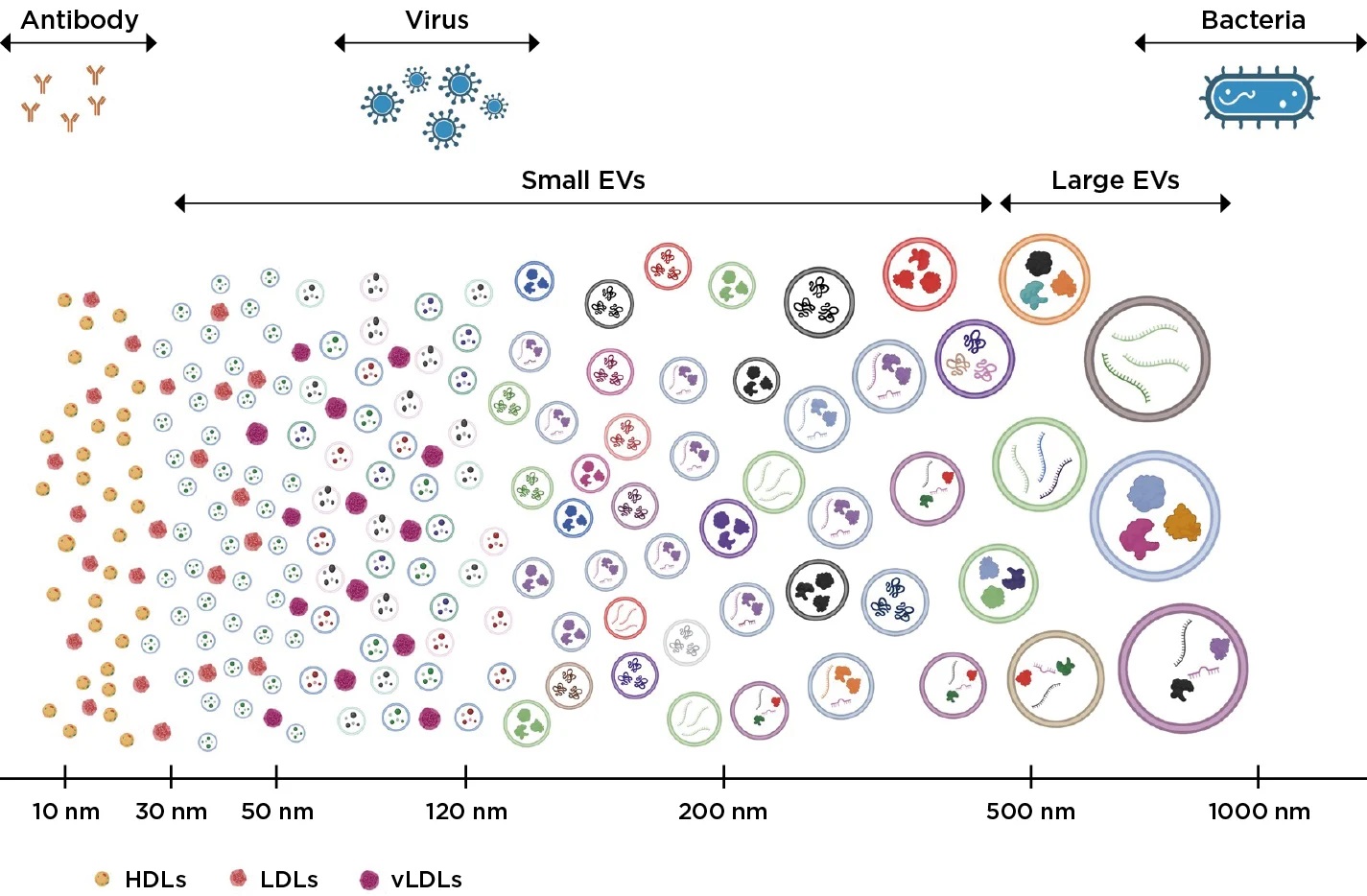
Source: Beckman Coulter
Figure 1. Size comparison of different cells and extracellular vesicles.
The CytoFLEX Nano Flow Cytometer can detect, characterize, and size extracellular vesicles as small as 40 nm.
Key Advantages and Features
Sensitivity and Multi-Channel Detection
- Six fluorescence channels and five side scatter channels enable detailed analysis of even the smallest particles.
- Increased laser power and specially engineered optical excitation paths capture more light, enhancing analytical efficiency.
Data Purity and Contamination Minimization
- Automated cleaning tools rapidly detect and eliminate contamination, reducing sample loss to <1%.
- Daily quality control identifies noise caused by nano- and microbubbles and triggers automatic purging and cleaning.
Optimized Optical and Fluidic Systems
- Reduced sample and sheath fluid flow rates allow work with micro-samples while maintaining high accuracy.
- A small-volume piston pump ensures stable and accurate counting (>90%).
- Specialized sheath fluid combined with a variable 5 nm filter minimizes background noise.
Ease of Use
- The intuitive CytExpert software simplifies system setup and adapts easily to users of all experience levels.
- Process automation reduces time and streamlines sample preparation.
The Power of the CytoFLEX Nano — Count, Characterize, and Size in One Instrument
The CytoFLEX Nano integrates all essential nanoscale analysis capabilities into a single instrument, enabling particle counting, characterization, and size determination.
SIZING
Accurate particle sizing by flow cytometry requires calibration using a well-characterized reference particle combined with an appropriate calibration method.
Latex polystyrene particles are the preferred tool, as they are NIST-traceable and manufactured with high consistency.
With the calibration method provided to every CytoFLEX Nano user, sizing data can be reliably reported in absolute units, allowing method comparison and correlation.
COUNT
The most robust particle-counting approaches rely on precisely controlled volumetric sample delivery, rather than manual sample loading, which introduces variability.
The CytoFLEX Nano allows users to tailor sample handling so that the exact sample volume needed for the required results is used.
CHARACTERIZE
Characterizing nanoscale particles requires best-in-class sensitivity.
To achieve this, the CytoFLEX Nano features a completely redesigned optical and fluidic system, maximizing the detection of even minimal fluorescence signals across six detection channels driven by four collinear lasers.
Explore the Full Picture of Your EV Experiment with the CytoFLEX Nano Flow Cytometer
This purpose-built flow cytometer delivers enhanced sensitivity, consistent performance, and the flexibility needed to study your samples with confidence.
Sensitivity
With exceptional size sensitivity from 1 µm down to 40 nm* and 10 nm resolution**, the CytoFLEX Nano enables the detection of smaller EV populations and their low-abundance cargo.
Flexibility
Expand your research possibilities with 6 fluorescence channels and 5 side scatter channels. Collect comprehensive data with fewer constraints.
Maximum Consistency
Working with nanoscale particles requires contamination-free, robust instrument performance.
Rely on reproducible data with >90% volumetric counting accuracy and <1% carryover between samples, supported by automated QC controls.
Familiarity
Built on the same principles as the CytoFLEX platform, the CytoFLEX Nano features a similar footprint and the same intuitive software interface for a seamless user experience.
Interested in Advancing Your Extracellular Vesicle Research?
Discover how the Beckman Coulter CytoFLEX Nano Flow Cytometer pushes beyond previous detection limits, providing researchers with access to data that was once out of reach.
When Choosing a Flow Cytometry Instrument, Three Key Factors Matter
Counting, sizing, and characterizing extracellular vesicles (EVs) provides researchers with deeper insights into cellular and organ health as well as disease signatures. However, EVs are difficult to analyze due to their small size and inherent heterogeneity.
The Need for Standardization
Until now, scientists have relied on multiple techniques to accurately count, characterize, and determine the size of EVs. This often results in workflows that are time-consuming and lack repeatability, making it harder to generate consistent and reliable data.
Webinar: First Experiences with the CytoFLEX Nano
This webinar features Alfonso Blanco, Director of Flow at University College Dublin, who presents preliminary results obtained by researchers studying extracellular vesicles (EVs) using the CytoFLEX Nano Flow Cytometer.
The session explores the limits of current flow cytometry technologies and explains how the CytoFLEX Nano is designed to overcome these challenges through a robust and consistent system capable of standardizing results across laboratories.
Our expert speaker, Alfonso Blanco, discusses the advantages of combining fluorescence with multiple scatter parameters to enable deeper characterization of nanoparticles and extracellular vesicles.
Press Review: What They Say About Recent Advances in Extracellular Vesicle Research
In the article “Developments in Extracellular Vesicle (EV) Research Present a Promising Future”, published in Technology Network (July 2024), Matthew Goff, Product Manager at Beckman Coulter, discusses several key themes:
- Traditional challenges in EV research
- Emergence of new technologies
- The importance of workflow automation
- How nanoscale technology is accelerating EV research
“...With greater clarity in results, EVs enable advanced diagnostic, prognostic, theranostic, and therapeutic applications, while reducing the invasiveness of sample collection. Monitoring disease progression or developing customized therapeutic models is incredibly powerful, but it requires highly specific analytical methods. This level of specificity is now finally available to a wide range of researchers.
Updating this workflow is truly an exciting moment in research...”
Beckman Coulter Life Sciences Revolutionizes Nanoparticle Analysis With the Launch of the CytoFLEX Nano Flow Cytometer
Article published in Technology Network — March 28, 2024
“...The CytoFLEX Nano Flow Cytometer shatters the previous limits of detection capabilities, arming researchers with data they previously would have never been able to access. This can open new doors for researchers and enable breakthrough discoveries in our battle against a myriad of diseases.
Extracellular vesicles can be found everywhere, and now researchers finally have a tool to see what has always been right in front of them.
We listened to the needs of our customers to create an analytical crossroads by increasing sensitivity through enhanced fluidics, optics, and electronics...”
Understanding Extracellular Vesicles
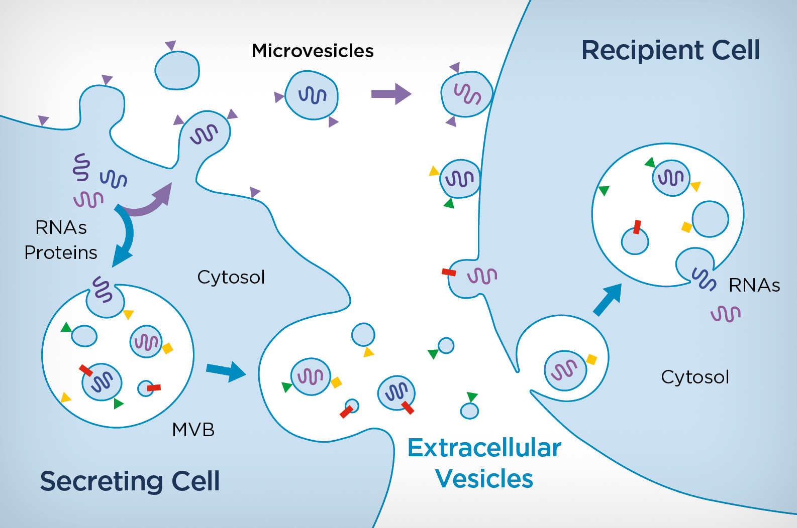
Extracellular vesicles (EVs) are small membrane-bound particles released by cells into the extracellular environment. They play essential roles in intercellular communication and are involved in numerous physiological and pathological processes. EVs are classified according to their biogenesis and size, primarily into exosomes, microvesicles, and apoptotic bodies.
Exosomes (Small EVs)
Exosomes typically range from 30 to 150 nm. They originate from the inward budding of multivesicular bodies (MVBs), which then fuse with the plasma membrane to release their content. Exosomes carry bioactive molecules—including proteins, lipids, nucleic acids (DNA, RNA), and microRNAs—that can be transferred to recipient cells and modulate their behavior.
Microvesicles (Large EVs)
Microvesicles, also referred to as large EVs, microparticles, or ectosomes, range from 100 to 1000 nm. Unlike exosomes, they are formed by outward budding and shedding of the plasma membrane. They transport a diverse cargo of proteins, lipids, and nucleic acids that can influence target cells.
Apoptotic Bodies
Apoptotic bodies are the largest EVs, ranging from 1 to 5 µm. They are released during apoptosis and contain cellular fragments, organelles, and nuclear material, which are cleared by phagocytic cells.
Growing Importance of EV Research
EVs have gained significant interest due to their potential as biomarkers for diagnosis and prognosis, their importance in cell-to-cell communication, and their possible therapeutic applications. Researchers analyze EVs in biological fluids such as blood, urine, and cerebrospinal fluid to better understand their cargo and functions.
Advanced technologies—including nano flow cytometry, electron microscopy, RNA sequencing, and proteomics—are essential for EV characterization. This growing field promises major advances in diagnostics, therapeutics, and regenerative medicine.
Current Challenges in EV Analysis
Despite their potential, EVs remain challenging to study:
- Isolation and purification methods often result in low yield, contamination, and limited standardization.
- EV populations are highly heterogeneous in size, cargo, and biogenesis.
- EVs exhibit diverse biological functions depending on their origin, making mechanistic interpretation complex.
- Functional heterogeneity requires improved characterization workflows and standardized assays.
One of the most significant limitations is the need to combine multiple techniques to accurately count, characterize, and size EVs. This leads to labor-intensive workflows, limited repeatability, and inconsistent results.