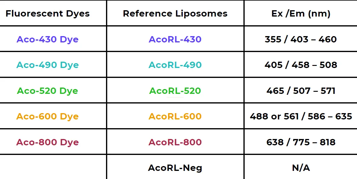
Acoerela: Innovative Optical Probes and Reference Standards for EVs, Nanoparticles, Bacteria, and Tumor Research
Traditional extracellular vesicle (EV) probes often produce non-specific signals, making data interpretation difficult.
Acoerela’s probes solve this issue by distinctly separating true biological signals from background noise. Their Reference Liposomes further support instrument optimisation and assay standardisation, enabling reproducible and reliable results.
These water-soluble membrane dyes offer highly specific and stable staining of cellular membranes with minimal background. They are suitable for a wide range of applications in imaging and flow cytometry.
Watch this one-minute video to discover Acoerela’s full range of advanced dyes.

The World's First Full Bilayer-Spanning Lipophilic Dyes
Water-Soluble Acoerela Dyes
Many conventional dyes are highly hydrophobic and require reconstitution in organic solvents such as DMSO or ethanol. However, these solvents can introduce artifacts in biological samples because they are less biocompatible than aqueous buffers.
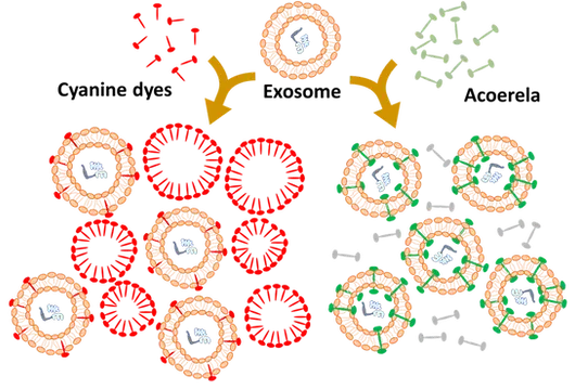
In addition, certain dyes—such as carbocyanines—tend to form nanoaggregates when serially diluted from DMSO or ethanol stock solutions. These nanoaggregates can generate false positive signals, particularly when analysing small biological particles such as exosomes.
Why Choose Acoerela
Fluorogenic Performance
Acoerela dyes exhibit a high quantum yield (>25%). A strong fluorescence increase is observed only when the dyes are bound to their target, due to changes in the local hydrophobic environment. The clear difference between bound and unbound emission profiles forms the basis of their “light-on” mechanism.
Diverse Color Options
The Acoerela chromophore platform currently supports up to three distinct colors. Ongoing developments aim to broaden the excitation and emission profiles, enabling expanded multiplexing capabilities across various applications.
Water Solubility
Acoerela dyes are highly water soluble and do not form micelles or nanoparticles when reconstituted in aqueous buffers. In contrast, dyes such as PKH26, DiR, and DiD can produce false positives in flow cytometry analyses of exosomes due to nanoparticle formation.
> 95 % Accuracy
Existing dyes exhibit false positivity rates exceeding 70%, whereas Acoerela dyes achieve >95% accuracy.
Fast:1 Hour
Current dyes may require more than eight hours to produce results. Acoerela dyes deliver reliable outcomes in approximately one hour.
Easy to Use: 1 Step
Most conventional dyes require multistep processing. Acoerela dyes streamline workflows with a simple one-step protocol.
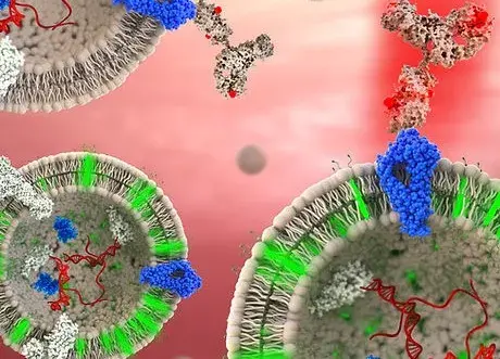
Extracellular Vesicle Dyes: Water-Soluble Dyes for Labeling Nanometer-Sized Vesicles and Exosomes
The highly water-soluble Acoerela Extracellular Vesicle Dyes enable accurate detection of nanosized extracellular vesicles, exosomes, and liposomes without artifacts or false positive signals originating from the dye itself. This performance is driven by Acoerela’s patented chemistry, which prevents self-assembly into micelles or nanoparticles.
Acoerela dyes are soluble in aqueous buffers (e.g., water, PBS, saline) at concentrations above 2 mg/mL. They exhibit strong emissive properties and fluoresce only when bound to their target. The dyes have been validated using exosomes derived from MSC, PC3, HT29, and A549 cell lines.
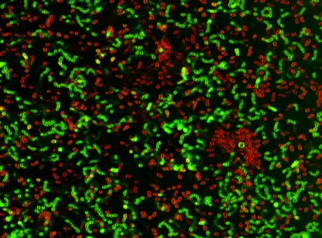
Gram-Typing of Bacteria
Acoerela offers dyes that enable visualization of Gram types in mixed bacterial populations without antimicrobial effects. Bacteria are typically classified as Gram-positive or Gram-negative based on their membrane structure. Gram-negative bacteria possess an outer membrane enriched with highly charged lipopolysaccharides (LPS), a feature absent in Gram-positive species.
The Acoerela Gram-Selective Dye leverages these structural differences and selectively labels Gram-positive bacteria due to the dye’s more rapid entry into their cells. Selectivity has been demonstrated in mixed cultures containing Gram-positive species (Staphylococcus aureus, Enterococcus faecalis) and Gram-negative species (Pseudomonas aeruginosa, Escherichia coli).
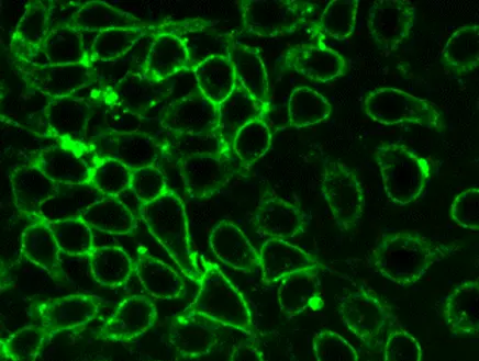
Mammalian Cell Membrane Dyes: Non-Cytotoxic Dyes for Tracking Cell Division Over Multiple Generations
Monitoring cell division is essential in cell biology research focused on immune regulation, cell function, quiescence, proliferation, and differentiation. Fluorescent cell division trackers are widely used in flow cytometry and imaging to study cell interactions and lineage outcomes both in vitro and in vivo.
Acoerela Membrane Dyes enable reliable tracking of mammalian cell division thanks to their stable membrane binding and low cytotoxicity. Their high quantum efficiency supports long-term fluorescence monitoring across multiple cell generations.
NR-II Tumor Tracking Dye: Near-Infrared II Probe for In Vivo Monitoring of Tumor Growth
Near-infrared II (NIR-II) dyes have gained significant interest in recent years due to their deep tissue penetration, low autofluorescence, and minimal background signals within this spectral window.
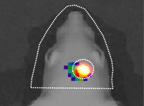
The Acoerela NIR-II dye enables long-term tracking of tumor growth in mouse models without loss of fluorescence intensity. Its durable emission and relatively high quantum yield allow researchers to monitor tumor progression and evaluate the effects of anticancer treatment regimens in vivo. This approach can reduce the number of test animals that would traditionally be sacrificed at regular intervals to assess tumor characteristics.
Extracellular Vesicles Dyes
Key Features
- Water soluble (>2 mg/mL)
- Stable labeling (>24 h)
- No inter-vesicle dye exchange
- Short incubation time (~30–60 min)
- Multiple color options available
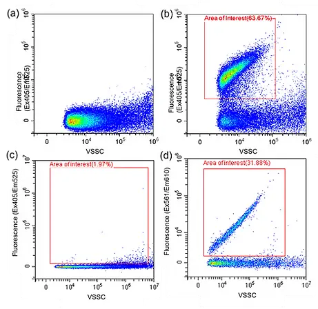
Flow Cytometry Analysis of Exosomes Labeled with Acoerela Extracellular Vesicle Dye
Dot plot showing:
(a) unstained exosomes,
(b) exosomes labeled with 1 µM Acoerela Exosome Dye,
(c) 1 µM Acoerela Exosome Dye alone, and
(e) 1 µM of a commercially available PKH26 dye alone, i.e., in the absence of exosomes.
Imaging Flow Cytometry
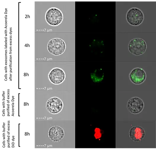
PC3 exosomes were labeled with Acoerela Extracellular Vesicle Dye and purified using an Amicon 100 kDa filter before being added to A549 cells, incubated, and analyzed on the ImageStream (Amnis) system after (a) 2 h, (b) 4 h, and (c) 8 h of incubation.
Acoerela-labeled exosomes were detected as green fluorescent spots.
In the absence of exosomes, no cellular fluorescence was observed because all free dye was removed during purification (d).
In contrast, DiD dye micelles were not removed, resulting in detectable fluorescence inside the cells. This signal reflects uptake of dye aggregates, which may be misinterpreted as DiD-labeled exosomes.
Bacteria Gram Selective Dyes
>GO TOP
Key Features
- Highly emissive
- Non-antimicrobial toward bacterial cells
- Suitable for both planktonic cultures and biofilms
- Excess dye is easy to wash off
The Acoerela Bacteria Gram-Selective Dye exploits structural differences between Gram-positive and Gram-negative cell walls. Because the dye enters Gram-positive bacteria more rapidly, it enables selective labeling of these species. Selectivity has been demonstrated in mixed cultures containing Gram-positive bacteria (Staphylococcus aureus, Enterococcus faecalis) and Gram-negative bacteria (Pseudomonas aeruginosa, Escherichia coli).
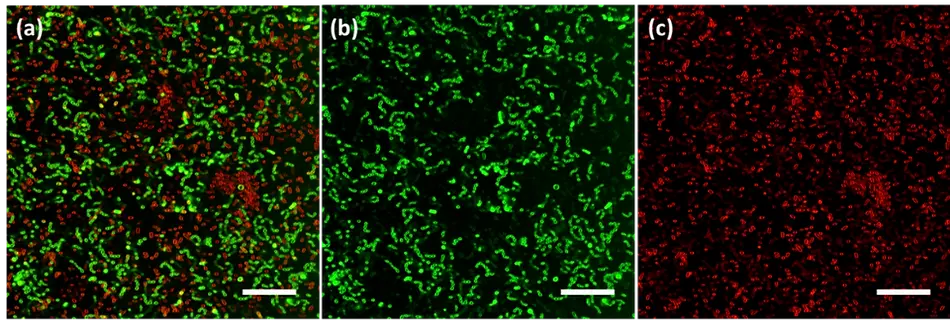
In situ Gram-typing of a mixed microbial biofilm was performed using the Acoerela dye together with a counterstain. Confocal microscopy images show E. faecalis (Gram-positive, coccus-shaped) and E. coli (Gram-negative, rod-shaped) in a polymicrobial biofilm after sequential staining with a Gram-positive–selective dye and a Gram-negative counterstain.
(A) Merged images from both channels:
(b) Acoerela Gram-Selective Dye channel (Ex405/Em450–490) and
(c) FM 4-64 channel (Ex639/Em640–700).
Membrane Dyes for Mammalian Cells > GO TOP
Monitoring cell division is essential in cell biology research focused on immune regulation, cell function, quiescence, proliferation, and differentiation. Fluorescent cell division trackers are widely used in flow cytometry and imaging to study cellular interactions and lineage outcomes both in vitro and in vivo.
Acoerela Membrane Dyes support precise tracking of mammalian cell division thanks to their stable membrane binding and low cytotoxicity. Their very high quantum yield enables long-term fluorescence monitoring across multiple generations.
Key Features
- High quantum yield (>25%)
- Stable binding to mammalian cell membranes
- Low photobleaching
- Low cytotoxicity
- Suitable for cell division tracking and red blood cell labeling
- Multiple color options available
Acoerela Cell Membrane Dye
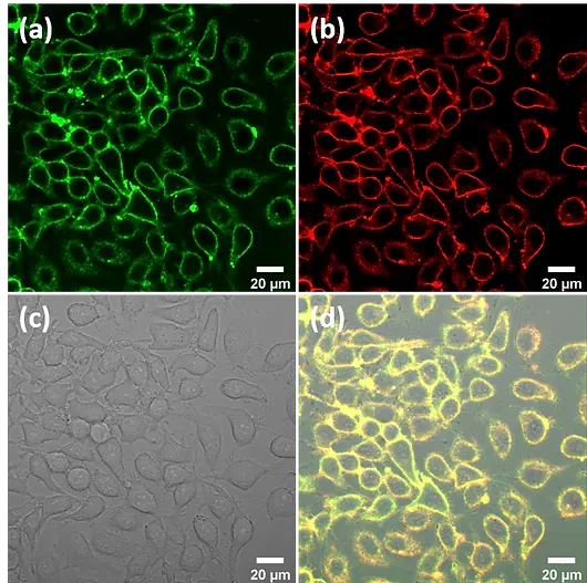
Confocal images of HepG2 cells co-stained with the Acoerela Membrane Labeling Dye and a commercially available membrane dye (FM 4-64).
(a) Acoerela dye channel (Ex405),
(b) FM 4-64 channel (Ex488),
(c) brightfield channel,
(d) merged channel.
The co-localization of Acoerela Membrane Dye with FM 4-64 confirms the membrane specificity of the Acoerela dye.
Acoerela Cell Division Tracking
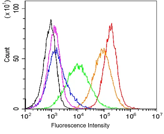
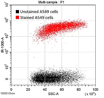
A549 mammalian tumor cells were labeled with the Acoerela Cell Division Tracking Dye. Labeled cells (red) showed a 3-log fold increase in fluorescence intensity compared with unstained cells (black). Fluorescence was maintained over four passages through serial passaging before analysis on the CytoFLEX (Beckman Coulter).
Orange: 1st passage, Green: 2nd passage, Blue: 3rd passage, Magenta: 4th passage.
Red Blood Cell Labeling
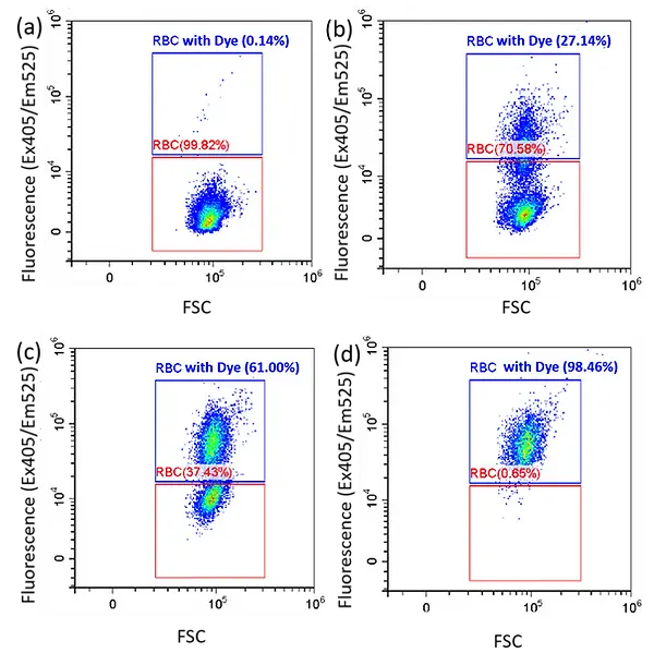
Flow cytometry analysis was performed on samples containing mixtures of unstained red blood cells (RBCs) and RBCs labeled with the Acoerela dye, at ratios of (a) 0:5, (b) 2:3, (c) 3:2, and (d) 5:0. The Acoerela RBC dye remains stable within the cells after labeling and does not transfer to unlabeled cells, ensuring clear and contamination-free population separation.
NIR-II Acoerela Dye > GO TOP
Near-infrared II (NIR-II) dyes have gained significant interest in recent years due to their ability to penetrate deeply into tissues and their low autofluorescence and background signal within this region of the electromagnetic spectrum.
The Acoerela NIR-II dye enables long-term tracking of tumor growth in mouse models without notable loss of emission. Its durable fluorescence and relatively high quantum yield allow researchers to monitor tumor progression and evaluate the impact of anticancer treatment regimens in vivo. This approach may reduce the number of test animals traditionally sacrificed at regular intervals for tumor characterization.
Acoerela Cell Membrane Dye
- Strong NIR-II fluorescence (Ex859/Em1017)
- Enables in vivo imaging
- Long-lasting signal (>14 days)
- Low cytotoxicity
- Highly water soluble
In Vivo Imaging
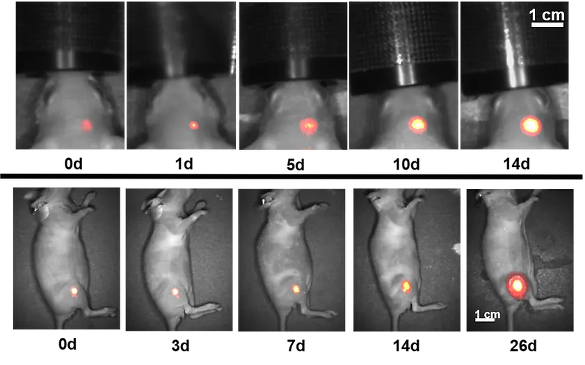
(Top) In vivo fluorescent images of an intracranial tumor over 14 days and (bottom) a subcutaneous tumor over 26 days, collected using emission from the NIR-II Acoerela Dye. In both cases, fluorescence intensity increased as the tumors grew, enabling non-invasive monitoring of tumor progression without the need for repeated sacrifice of the mice.
AcoRL Reference Liposomes
AcoRL Reference Liposomes are pre-labeled fluorescent liposomes formulated with biologically relevant synthetic lipids and 1 mol% Aco-Dyes (relative to total lipid content). They function as reliable standards for instrument optimisation and for supporting fluorescence-based analyses.
Available as both positive and negative controls, AcoRL liposomes have an average particle size of 50–100 nm. They enable accurate visualization and analysis of Aco-Dye–labeled samples using flow cytometry or other fluorescence-based platforms.
ASK FOR A QUOTE : ANALIS - NAMUR 081 25 50 50 - GENT 092437710 - WWW.ANALIS.COM/ACOERELA - MAIL@ANALIS.BE